Contrary to common belief, the greatest advantage of digital X-rays isn’t just speed, but their power to make the nearly invisible, visible.
- Digital systems reveal subtle hairline fractures and bone changes that traditional film often misses entirely.
- They enable a data-driven approach, turning a radiologist’s interpretation from a simple observation into a calculated probability.
Recommendation: When discussing results, focus on the diagnostic confidence level the technology provides, not just the image itself.
As a radiologist, I’ve spent countless hours in darkened rooms, my eyes tracing the faint grey and white landscapes of the human body. For decades, the gold standard was film—a physical, tangible piece of evidence. We were trained to spot the obvious breaks, the clear dislocations. But the real challenge, the one that keeps us on our toes, has always been the subtle, the ambiguous, the hairline fracture that masquerades as normal anatomy. Patients and colleagues often assume the main benefit of modern digital radiography is speed or lower radiation. While true, these are just convenient byproducts.
The real revolution isn’t logistical; it’s diagnostic. The conversation has shifted from « Is it broken? » to « What is the statistical probability that this faint line is a fracture? » We’ve moved beyond simple observation into the realm of data analysis. Digital imaging allows us to manipulate contrast, zoom into micro-textures, and even use algorithms to flag areas the human eye might glide over. This isn’t just about getting a « clearer picture. » It’s about enhancing our perception and quantifying our uncertainty.
But this technological leap introduces new questions. What do we do with unexpected findings, like a tiny lung nodule? When does the quest for more certainty, via a CT scan, justify a higher radiation dose? And how do we, as humans, work with artificial intelligence without falling prey to our own cognitive biases? This article explores not just how digital X-rays are better, but how they have fundamentally changed the science of seeing inside the body, empowering us to catch what was once missed.
To fully grasp this evolution in diagnostic imaging, we will explore the nuances that differentiate modern techniques from their predecessors. The following sections break down key scenarios where digital technology’s impact is most profound, from identifying incidental findings to collaborating with artificial intelligence.
Summary: The New Era of Diagnostic Precision
- What Does « Incidental Lung Nodule » Mean and Should You Worry?
- How Often Should You Repeat X-rays for Monitoring Osteoporosis?
- X-Ray vs CT Scan: When Is More Radiation Justified?
- The « Normal » X-Ray That Missed Hairline Fracture Until 6 Weeks Later
- How to Position Yourself During X-Rays to Get Clear Images on First Attempt
- Radiologist vs AI Algorithm: Who Spots Pneumonia First?
- The Correlation Mistake: Why Eating Almonds Doesn’t Cause Longevity
- Why 95% Confidence Intervals Matter More Than P-Values
What Does « Incidental Lung Nodule » Mean and Should You Worry?
An « incidental lung nodule » is a small, spot-like shadow on the lung that is discovered unintentionally, usually during a chest X-ray performed for another reason, like a suspected rib fracture. It’s the perfect example of visual ambiguity. Most of these nodules are benign, representing old scars from past infections or other harmless tissue. However, the initial image doesn’t tell the whole story, and the primary role of the radiologist is to determine the likelihood of it being something more serious, without causing undue alarm.
This is where digital technology provides a significant advantage. Instead of a static film image, we have dynamic data. We can adjust brightness and contrast to better define the nodule’s edges and density. The ability to easily compare with prior digital images is also crucial for tracking stability or growth over time. Furthermore, the rise of AI is transforming our ability to even detect these findings. For instance, a 2023 study found that AI could detect incidental lung nodules on chest X-rays in about 1.0% of patients in a large cohort, a discovery rate that is difficult to achieve with human review alone under high-volume conditions.
The key is not to panic. An incidental finding is a starting point for a conversation, not an immediate diagnosis. Research has shown that while many nodules are found, only a fraction require intervention. In fact, one analysis revealed that of all the incidental nodules detected with AI assistance, only about 20.6% required clinical management, including cases of malignancy or active inflammation. The initial report is a probabilistic diagnosis, and follow-up imaging or consultation is the standard, logical next step to gain more certainty.
How Often Should You Repeat X-rays for Monitoring Osteoporosis?
Monitoring chronic conditions like osteoporosis presents a classic medical dilemma: the need for regular imaging to track disease progression versus the desire to minimize cumulative radiation exposure. While a standard X-ray can show fractures that are a consequence of osteoporosis, the primary tool for measuring bone mineral density (BMD) is a specialized, low-dose scan called a DXA (or DEXA) scan. These scans provide the quantitative data needed to diagnose and monitor the condition itself, with studies confirming the patient dose from DXA is extremely low, often comparable to a day’s worth of natural background radiation.
However, X-rays still play a vital role, particularly in assessing for vertebral compression fractures, which can be silent and are a hallmark of advancing osteoporosis. This is where digital radiography’s efficiency shines. The ability to perform repeated imaging over years is made more acceptable because modern digital X-ray systems achieve a 30-80% reduction in radiation exposure compared to old film-based systems. This significant reduction allows clinicians to order follow-up images with greater confidence when a patient reports new back pain or height loss.
The frequency of repeat imaging is not one-size-fits-all. It depends on several factors: the patient’s baseline BMD, their rate of bone loss, treatment status, and the presence of new symptoms. Typically, a follow-up DXA scan is recommended every one to two years after initiating treatment. X-rays are performed on an as-needed basis when a new fracture is suspected. The digital archive is invaluable here, allowing for instantaneous side-by-side comparison with previous images to detect subtle changes in vertebral height or shape, a task that was cumbersome and less precise with physical film records.
X-Ray vs CT Scan: When Is More Radiation Justified?
A standard X-ray is a two-dimensional shadow of a three-dimensional object. For many simple fractures, this is sufficient. But what happens when a fracture is complex, involves a joint, or is suspected but not visible on the X-ray? This is when we consider a Computed Tomography (CT) scan. A CT scan is essentially a series of X-rays taken from multiple angles, which a computer then reconstructs into detailed cross-sectional images. It eliminates the problem of overlapping structures, providing an unparalleled view of bone and soft tissue.
This clarity comes at a cost: a higher radiation dose. As a radiologist, my guiding principle is « ALARA » – As Low As Reasonably Achievable. We do not order a CT scan lightly. The justification hinges on a crucial question: Will the additional information gained from the CT scan fundamentally change the patient’s treatment plan? If an X-ray shows a wrist fracture that clearly requires a cast, a CT is often unnecessary. But if the fracture involves the intricate carpal bones, or if we suspect a subtle fracture in the spine or pelvis, a CT is essential for surgical planning and preventing long-term complications. The dose difference is not trivial; research on radiation exposure shows that a volumetric CT can deliver 1-3 mSv, while a digital radiograph’s dose is significantly lower.
Emerging technologies like digital tomosynthesis are helping to bridge this gap. This technique acquires multiple low-dose projections at different angles to create a series of slices, offering better depth perception than a single X-ray without the full radiation dose of a CT.

As this visualization suggests, tomosynthesis allows us to digitally « peel away » layers, helping to resolve ambiguous findings and reduce the need for an immediate jump to CT. The decision is always a careful balance, weighing the diagnostic benefit against the radiation risk. The added certainty from a CT is justified only when it directly informs a critical treatment decision that an X-ray cannot.
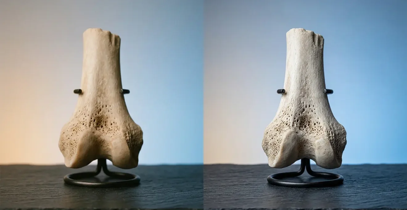
The « Normal » X-Ray That Missed Hairline Fracture Until 6 Weeks Later
One of the most common and frustrating scenarios in orthopedics is the « occult » fracture—a fracture that is truly present but invisible on the initial X-ray. The scaphoid, a small bone in the wrist, is notorious for this. A patient falls on an outstretched hand, has classic point tenderness, but the X-ray comes back « normal. » The patient is told it’s a sprain, but the pain persists. Weeks later, a repeat X-ray shows a clear fracture line, often with signs of poor healing. This delay can lead to serious long-term consequences like non-union or avascular necrosis.
This is not necessarily an error of interpretation; it’s a limitation of the technology. On a film X-ray, a non-displaced hairline fracture can be completely obscured by the overlapping shadows of other bones. This is a prime example of low « signal-to-noise ratio. » The fracture (signal) is too faint to be distinguished from the background anatomy (noise). In fact, clinical research demonstrates that the initial detection rate for scaphoid fractures on radiographs is only around 70%, meaning nearly a third are missed.
This is where digital imaging provides its most profound advantage. The ability to manipulate the digital data is a game-changer. We can zoom in on the area of concern, apply edge-enhancement algorithms, and adjust the window and level (contrast and brightness) to make a faint line « pop. » This post-processing capability significantly increases the sensitivity for detecting these subtle injuries. An illustrative study highlights this gap perfectly.
Case Study: Digital Imaging vs. Conventional Radiography for Occult Fractures
A 2015 study of 124 patients with suspected scaphoid fractures revealed a stark contrast in diagnostic accuracy. Conventional radiography detected only 42.8% of the fractures that were ultimately confirmed. In 24 of these patients, the initial film X-ray was interpreted as completely normal, showing no fracture. However, subsequent advanced imaging with multidetector CT, a digital modality, clearly demonstrated the fracture line. This highlights how hairline fractures can remain completely invisible on static film but become clearly visible with the advanced contrast resolution and post-processing capabilities of digital imaging technologies.
This is the core of the digital promise: not just to see the obvious better, but to reveal the ambiguous and prevent the diagnostic delays that can compromise patient outcomes.
How to Position Yourself During X-Rays to Get Clear Images on First Attempt
While the radiologist’s interpretation is critical, the quality of a diagnostic image begins much earlier: with the technologist and the patient in the X-ray room. A poor-quality image, whether due to patient movement or incorrect positioning, is the primary reason for needing retakes. Each retake means additional radiation exposure and a longer time spent in the clinic. The goal is always to get the perfect shot on the first attempt, and digital X-rays have revolutionized this process through one key feature: the immediate feedback loop.
With old film systems, the technologist would position the patient, take the exposure, and then have to take the film cassette to a darkroom for processing—a process that could take several minutes. Only then would they know if the image was adequate. In contrast, digital radiography produces viewable images within 3-5 seconds on a monitor right in the exam room. This instant review allows the technologist to confirm image quality before the patient even moves. If a subtle adjustment is needed, it can be done on the spot.
As a patient, you play an active role in this process. The single most important thing you can do is to hold perfectly still during the brief moment of exposure. Listen carefully to the technologist’s instructions, as they are designed to place the area of interest in the optimal position, free from overlying structures. Relaxing your muscles and holding your breath when asked can make a significant difference in image sharpness. The following checklist, used by technologists, is made possible by the speed of digital imaging.
Action Plan: Achieving a First-Attempt-Success X-ray
- Immediate Verification: The technologist reviews the digital image on their monitor seconds after exposure to check for motion or positioning errors while you are still in position.
- Real-Time Quality Control: They can instantly assess if the entire area of interest is captured and if the angle is correct, preventing the need for you to be called back.
- Software Optimization: Minor exposure issues can often be corrected with software adjustments to contrast and brightness, saving a retake for a slightly over- or under-exposed image.
- Minimize Movement: The technologist confirms a successful image before you are asked to move, which is the most critical step to avoid a repeat exposure.
- Secure Archiving: The image is instantly and electronically archived, eliminating the risk of a physical film being lost, which would necessitate a complete recall and re-exposure.
This streamlined workflow not only improves efficiency and safety but also ensures the radiologist receives the highest quality data possible for an accurate diagnosis.
Radiologist vs AI Algorithm: Who Spots Pneumonia First?
The question of « man vs. machine » in radiology is a popular topic, but it sets up a false dichotomy. The future—and indeed, the present—is not about replacement, but about collaboration. Artificial intelligence, specifically deep learning algorithms, are incredibly powerful tools for pattern recognition. They can be trained on millions of images to spot subtle abnormalities that might indicate conditions like pneumonia or, more relevant to our topic, fractures.
So, who is faster? An AI can screen a chest X-ray in milliseconds, flagging potential areas of concern far quicker than a human can. However, speed is not the same as accuracy or understanding. An AI algorithm might flag a shadow, but it doesn’t understand the patient’s clinical history, symptoms, or the myriad of other factors that contribute to a final diagnosis. This is where the radiologist’s expertise is irreplaceable. My role is evolving from a primary detector to an expert verifier and integrator of data. The AI acts as a second pair of eyes—a tireless, hyper-vigilant assistant.
This collaborative approach demonstrably improves outcomes. The AI provides a « first pass, » drawing my attention to potential findings and increasing my diagnostic sensitivity. For fracture detection, the results are compelling. A major 2022 Radiology study found a 10.4% improvement in sensitivity for fracture detection when radiologists used AI assistance compared to reading on their own. The AI helps reduce perceptual errors and ensures that subtle findings aren’t overlooked during a busy day.

This concept of augmented perception is the correct way to view this partnership. The AI provides quantitative analysis and flags probabilities, while the human expert provides context, clinical correlation, and ultimate judgment. It’s not about who spots it « first, » but how we can combine our strengths to arrive at the most accurate and timely diagnosis for the patient.
Key Takeaways
- The true value of digital imaging is not speed but diagnostic precision and the ability to quantify uncertainty.
- Digital tools and AI act as an extension of the radiologist’s perception, helping to detect subtle findings that film can miss.
- An effective diagnosis is a collaborative process involving technology, the technologist, the radiologist, and the patient.
The Correlation Mistake: Why Eating Almonds Doesn’t Cause Longevity
In the age of big data and AI, we have an unprecedented ability to find patterns. An algorithm can analyze millions of data points and tell us that people who eat almonds tend to live longer. This is a correlation. The critical error, both in public understanding and sometimes in clinical practice, is to assume this means almonds *cause* longevity (causation). In reality, people who eat almonds might also be more likely to exercise, have better access to healthcare, and engage in other healthy behaviors. The almonds are a marker, not the cause.
This same cognitive trap exists in radiology, even with our advanced digital tools. We might observe that patients with a certain type of faint shadow on their X-ray tend to develop arthritis five years later. Is the shadow causing the arthritis, or is it merely an early, non-specific marker of an underlying process that causes both? This distinction is paramount. As a radiologist, I must constantly resist the urge to over-interpret a correlation as a cause. The image is a piece of evidence, not a complete narrative.
Observer variability is another significant challenge that technology doesn’t entirely solve. Even with precise digital images, different radiologists may interpret the same subtle findings differently. This is the « honest » part of being a radiologist; we must acknowledge the inherent subjectivity in our work. As one seminal paper on the topic noted:
Many basic radiographic interpretations relied on in making treatment decisions are made variably by observers. Using experienced raters and precise definitions of fracture assessments does not guarantee a high level of agreement.
– Martin J, Marsh JL, et al., Journal of Orthopaedic Trauma, 2000
Digital imaging and AI help by standardizing measurements and flagging areas of interest, reducing some of this variability. However, the final interpretation still rests on human judgment, which must be disciplined to distinguish a meaningful signal from a coincidental correlation.
Why 95% Confidence Intervals Matter More Than P-Values
This brings us to the philosophical heart of modern diagnostics. For years, medicine was obsessed with the « p-value, » a statistical measure often misinterpreted as the probability of a finding being « true. » A more honest and useful concept, especially in imaging, is the confidence interval (CI). Instead of a simple « yes » or « no, » a 95% CI gives us a range of plausible values for a measurement. It’s a direct statement of our diagnostic precision.
Imagine measuring the gap in a fractured bone. A film X-ray might let me say, « the gap is about 2 millimeters. » With high-resolution digital imaging and measurement tools, I can say, « we are 95% confident that the true gap is between 1.9 and 2.1 millimeters. » This level of precision is vital for a surgeon deciding between a cast and a surgical plate. The confidence interval quantifies our uncertainty. A narrow CI means high confidence and a precise measurement; a wide CI tells us our measurement is less reliable and we should be more cautious.

This shift in thinking is profound. It moves us away from a binary world of « fractured » or « not fractured » and into a more realistic, probabilistic framework. The power of digital imaging is its ability to provide the data needed to calculate these narrow confidence intervals. When a modality like MRI is used for an occult scaphoid fracture, its diagnostic accuracy can be incredibly high. A landmark 1997 Radiology study reported sensitivity and specificity approaching 100%, which translates to an extremely narrow and reliable confidence interval around the diagnosis.
Ultimately, the goal of all this technology isn’t to produce a beautiful picture. It is to provide the most accurate and precise data possible, so that we can make a diagnosis with the highest, most quantifiable level of confidence. It’s about being honest about what we know, and more importantly, what we don’t know.
Therefore, the next time you see a digital X-ray, look beyond the image itself. See it as a rich dataset, one that allows medical professionals to move past simple observation and toward a more precise, data-driven, and confident diagnosis. To put these concepts into practice, the most important step is to engage in a detailed conversation with your physician about the certainty of your imaging results.